Living Light: The Mechanisms and Diversity of Bioluminescence
Living Light: The Mechanisms and Diversity of Bioluminescence
[EDITOR’S NOTE: The following article was co-authored by AP auxiliary staff scientist Dr. Joe Deweese and two of his talented undergraduate research assistants, Luke Sullivan and Caleb Hammond, both of whom are former Apologetics Press camp attendees. Dr. Deweese holds a Ph.D. in Biochemistry from Vanderbilt University and serves as Professor of Biochemistry and Director of Undergraduate Research at Freed-Hardeman University.]
Introduction
On a summer night, one of nature’s most captivating sights is the flickering of fireflies (Figure 1). These glowing insects, like many other organisms, exhibit bioluminescence—the ability to produce light through chemical reactions in their bodies. Bioluminescence can be found in various creatures, from land-based fungi to ocean-dwelling bacteria. In fact, approximately 76% of all marine organisms are bioluminescent.1

Bioluminescence occurs in three main ways. The first mechanism is the luciferin-luciferase system, which involves molecular oxygen, a light-producing molecule called luciferin, and an enzyme called luciferase. Luciferase helps oxygen react with luciferin, which leads to light production (Figure 2).2 While the structures and chemical compositions of luciferins and luciferases vary across species, many organisms, such as fireflies and marine plankton, use a form of this system (Figure 3).3


The second form of bioluminescence uses fluorescent proteins, which absorb one wavelength of light and emit another. While the green fluorescent protein (GFP) from jellyfish has greatly benefitted the scientific community, its mechanism is different from other luminescent proteins.4 The chromophore, the part that absorbs and re-emits light, is located in the primary structure of the protein, which depends upon its amino acid sequence.5 Researchers have modified GFPs to use in research applications, altering the wavelengths of light that are absorbed and emitted.
Finally, there are the photoproteins. These molecules are similar to luciferin in that they react with oxygen to emit light, but the reaction does not require a luciferase. Interestingly, photoproteins and GFP have been discovered as working together in jellyfish.6 It is unclear how widespread photoproteins are among living organisms.
In some cases, bioluminescence is found in organisms that do not generate the light but instead exist in a symbiotic relationship with bioluminescent bacteria. For example, the Anglerfish produces light through a specialized organ, called the esca, that houses bioluminescent bacteria.7 These bacteria use a luciferin-luciferase type system, but it is very distinct from that found in fireflies and other eukaryotic organisms. The remainder of this article aims to explore examples of the diversity of the luciferin-luciferase bioluminescent systems.
Diversity of Luciferin-Luciferase Systems
Luciferin-luciferase systems vary significantly across organisms. The term “luciferin” broadly refers to any molecule that reacts with oxygen to generate light. Examples of different luciferins are shown in Figure 4. Organisms must not only produce luciferin but also create enzymes to regulate its production and control the timing of light production.8

There is also great diversity in luciferases.9 Examples of bacterial and eukaryotic luciferases are shown in Figure 5. The amino acid sequences of these enzymes are highly diverse, illustrating that these enzymes are structurally unique across species.

Some evolutionary scientists suggest that luciferases evolved independently in different organisms. For example, the luciferase system in fireflies is believed to have evolved separately from the marine systems. Each system has unique biochemical pathways, supporting the idea that these systems are highly specialized.10
Bacterial Luciferin-Luciferase System
Though bacteria are simple organisms, their bioluminescent systems are complex. Bioluminescent bacteria contain genes that encode the proteins necessary for light production. Some evolutionists believe these genes evolved independently in many species, while others argue that they spread through horizontal gene transfer (i.e., the ability of some organisms to transfer genetic information to other organisms).11
In bacteria, two proteins, LuxA and LuxB, form a complex called LuxAB. This complex converts aldehydes into acids, using FMNH2 as an electron source. There is debate over the precise nature of the chemicals involved in the reaction, but long-chain aldehydes are generally accepted as the substrates.12 Several other reactants and genes regulate the light’s intensity and other aspects of the system.13 Despite the continued exploration of bacterial bioluminescence, much remains to be discovered about how all the components work together.14
Fungal Luciferin-Luciferase System
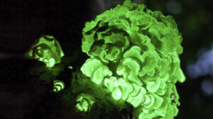
Bioluminescent fungi, which can be found worldwide, have fascinated scientists for centuries. Approximately 80 species of glowing fungi have been identified (see Figure 6 for an example). However, it was only in 2015 that the luciferin responsible for fungal bioluminescence was discovered. Researchers found that the molecule 3-hydroxyhispidin acts as luciferin in fungi, a molecule also found in some plants.15

Fungal bioluminescence seems to be a by-product of metabolism, as the light is constantly produced. The intensity of the light varies with the time of day, suggesting a connection to circadian rhythms.16
The process of light production in fungi involves two steps. First, a precursor molecule, hispidin, is converted into 3-hydroxyhispidin by an enzyme. This luciferin is then activated to produce light. Although researchers are still working to fully understand the genetic mechanisms behind this process, the general reaction has been established.17
Dinoflagellate Luciferin-Luciferase System
Dinoflagellates, tiny single-celled organisms that are a type of phytoplankton, produce one of the most stunning forms of bioluminescence and are responsible for the glowing ocean waters often observed at night (Figures 3 and 7). Unlike other systems, dinoflagellates produce light in specialized organelles called scintillons, which contain luciferin, luciferase, and sometimes a protein that binds to luciferin.18

The luciferin in dinoflagellates is structurally similar to chlorophyll, the pigment that helps plants photosynthesize. While the exact structure of luciferin in most dinoflagellates is still unknown, the luciferin from P. lunula has been studied extensively and is thought to react with the luciferase of other dinoflagellates. The entire system is regulated by the day-night cycle. In some species, scintillons are destroyed at dawn and recreated at dusk, while in others, the molecules are simply relocated to different parts of the cell.19 Dinoflagellates also have Luciferin Binding Protein (LBP), which is suspected to hold onto the luciferin under normal conditions and release it when the acidity of the cell changes. However, the exact function and mechanism is not known.20 Though much remains unknown about the function of dinoflagellate bioluminescence, it continues to intrigue scientists for its complexity and beauty.
Key Observations and Reflections
The diversity among bioluminescent organisms is remarkable, particularly within the luciferin-luciferase systems. Each organism has its own specific luciferin and luciferase, which are not interchangeable between different groups of organisms (e.g., bacterial luciferin cannot substitute for fungal luciferin). Furthermore, the luciferin-luciferase mechanism is dependent upon several components, including genetic information, light-producing luciferin, luciferase enzyme, oxygen, various cofactors, and environmental conditions. Considering the immense diversity and intricate complexity, we must ask a fundamental question: how did these organisms develop this ability to produce light? There are only two options: unguided evolution or intentional design.
Within the evolutionary framework, there are a few postulated possibilities. First, bioluminescence could have originated from an ancestral organism with a primitive luciferin-luciferase system, which evolved into the diverse systems we see today. However, the vast diversity between systems makes it difficult to attribute them all to a common ancestor.21 Thus, most evolutionary scientists propose that these systems evolved independently in different organisms multiple times!22 Again, however, this is problematic. The statistical probability of this complex system evolving one time is low enough; the likelihood of these mechanisms arising across all bioluminescent organisms is practically null.
Therefore, intentional design is the best explanation for the origin of this diverse, complex phenomenon. And because that Designer must be supernatural in order to invent nature, and because the Bible must be the work of a perfect Being in Whom is no flaw, we can humbly and confidently join the Psalmist in professing that all of creation is “the work of [the LORD’s] fingers,” including this fascinating mechanism of bioluminescence (Psalm 8:3).23 Furthermore, let’s praise Him because the same God who designed the glowing dinoflagellates is “mindful” of us (v. 4). As verse 9 so beautifully says, “O LORD, our Lord, how excellent is Your name in all the earth!”
Conclusion
Bioluminescence represents a diverse set of mechanisms in a broad spectrum of organisms. Even within the mechanisms known as “luciferin-luciferase” systems, the degree of diversity is staggering. The natural origin of these mechanisms would require multiple evolutionary miracles in order to develop the genes and control mechanisms needed to carry out these functions. These complex systems and their elaborate control systems are best explained by means of a supernatural Creator.
Endnotes
1 Séverine Martini and Steven H.D. Haddock (2017), “Quantification of bioluminescence from the surface to the deep sea demonstrates its predominance as an ecological trait,” Scientific Reports, 7:1-11, April.
2 Osamu Shimomura (2012), Bioluminescence: Chemical Principles and Methods (Singapore: World Scientific), revised edition.
3 Ibid.
4 Osamu Shimomura, Frank H. Johnson, and Yo Saiga (1962), “Extraction, Purification and Properties of Aequorin, a Bioluminescent Protein from the Luminous Hydromedusan, Aequorea,” Journal of Cellular and Comparative Physiology, 59[3]:223-239, June; Osamu Shimomura (1979), “Structure of the chromophore of Aequorea green fluorescent protein,” FEBS Letters, 104[2]:220-222, August; Shimomura (2012); Sowmya Swaminathan (2009), “GFP: the green revolution,” Nature Cell Biology, 11[1]:S20, October.
5 Shimomura (1979).
6 Shimomura (1962).
7 Lindsay L. Freed, Cole Easson, Lydia J. Baker, Danté Fenolio, Tracey T. Sutton, Yasmin Khan, Patricia Blackwelder, Tory A. Hendry, and Jose V. Lopez (2019), “Characterization of the microbiome and bioluminescent symbionts across life stages of Ceratioid Anglerfishes of the Gulf of Mexico,” FEMS Microbiology Ecology, 95[10]:1-11, September.
8 Eveline Brodl, Andreas Winkler, and Peter Macheroux (2018), “Molecular Mechanisms of Bacterial Bioluminescence,” Computational and Structural Biotechnology Journal, 16:551-564, November; David Morse, A.M. Pappenheimer, Jr., and J. Woodland Hastings (1989), “Role of a luciferin-binding protein in the circadian bioluminescent reaction of Gonyaulax polyedra,” Journal of Biological Chemistry, 264[20]:11822-11826, July; Konstantin V. Purtov, Valentin N. Petushkov, et al. (2015), “The Chemical Basis of Fungal Bioluminescence,” Angewandte Chemie International Edition, 54[28]:8124-8128, July; R.L. Airth and G. Elizabeth Foerster (1962), Archives of Biochemistry and Biophysics, 97[3]:567-573, June.
9 Shimomura (2012).
10 Aubin Fleiss and Karen S. Sarkisyan (2019), “A brief review of bioluminescent systems,” Current Genetics, 65:877-882, March; Asiri N. Dunuweera, Shashiprabha P. Dunuweera, and K. Ranganathan (2024), “A Comprehensive Exploration of Bioluminescence Systems, Mechanisms, and Advanced Assays for Versatile Applications,” Biochemistry Research International, 1-22, February; Thérèse Wilson, and J. Woodland Hastings (1998), “Biolumiscence,” Annual Review of Cell and Developmental Biology, 14:197-230, November.
11 Eveline Brodl, Andreas Winkler, and Peter Macheroux (2018), “Molecular Mechanisms of Bacterial Bioluminescence,” Comput Struct Biotechnol J., November. For a response, see Joe Deweese (2015), “What Is Horizontal Gene Transfer, and Does It Support Evolution?” Reason & Revelation, 35[9]:100-105, September.
12 Ibid.
13 Ibid; Edward Meighen (1989), “Bacterial bioluminescence: organization, regulation, and application of the lux genes,” Federation of American Societies for Experimental Biology Journal, 7[11]:1016-1022, August.
14 Brodl, Winkler, and Macheroux (2018).
15 Audrey Chew, Dennis Desjardin, Yee-Shin Tan, Md Yusuf Musa, and Vikineswary Sabaratnam (2014), “Bioluminescent Fungi from Peninsular Malaysia—a Taxonomic and Phylogenetic Overview,” Fungal Diversity, September; Konstantin Purtov, Valentin Petushkov, et al. (2015), “The Chemical Basis of Fungal Bioluminescence,” Angewandte Chemie International Edition, 54(28):8124-8128, July.
16 Konstantin Purtov, Valentin Petushkov, et al. (2015), “The Chemical Basis of Fungal Bioluminescence,” Angewandte Chemie International Edition, 54[28]:8124-8128, July; Anderson Oliveira, Cassius Stevani, et al. (2015), Current biology, 25[7]:964-968, March.
17 Ibid; R.L. Airth and G. Elizabeth Foerster (1962), “The Isolation of Catalytic Components Required for Cell-Free Fungal Bioluminescence,” Archives of Biochemistry and Biophysics, 97:567-573, January; Alexey Kotlobay, Karen Sarkisyan, et al. (2018), “Genetically encodable bioluminescent system from fungi,” Proceedings of the National Academy of Sciences of the United States of America, 115[50]:12728-12732, November; Kseniia Palkina, Anastasia Balakireva, et al. (2023), “Domain Truncation in Hispidin Synthase Orthologs from Non-Bioluminescent Fungi Does Not Lead to Hispidin Biosynthesis,” International Journal of Molecular Sciences, 24[2]:1317, January.
18 Martha Valiadi and Debora Iglesias-Rodriguez (2013), “Understanding Bioluminescence in Dinoflagellates—How Far Have We Come?,” Microorganisms, 1,1 3-25.5, September.
19 Michel Desjardins and David Morse (1993), “The Polypeptide Components of Scintillons, the Bioluminescence Organelles of the Dinoflagellate Gonyaulax Polyedra,” Biochemistry and Cell Biology, 71[3-4]:176-182, March; Valiadi and Iglesias-Rodriguez (2013).
20 Valiadi and Iglesias-Rodriguez (2013); D. Morse, A.M. Pappenheimer Jr, and J.W. Hastings (1989), “Role of a luciferin-binding protein in the circadian bioluminescent reaction of Gonyaulax polyedra,” The Journal of Biological Chemistry, 264[20]:11822-11826, July.
21 Shimomura (2012)
22 Fleiss and Sarkisyan (2019).
23 Eric Lyons and Kyle Butt (2014), “7 Reasons to Believe in God,” Reason & Revelation, 34[10]:110-119, October.

REPRODUCTION & DISCLAIMERS: We are happy to grant permission for this article to be reproduced in part or in its entirety, as long as our stipulations are observed.


